Modern medical imaging and technology have vastly improved and enhanced healthcare.
Modern medical imaging techniques, such as MRI, CT, ultrasound, and X-ray, are helping to identify serious life-threatening disorders, diseases, and injuries, and, in this way, have saved millions of lives around the world.
However, where modern imaging fails is in how it has been misused to explain the pain. It is still common knowledge that pain is caused by a structural anomaly seen on a scan e.g. a degenerative change or an irregularity to the so-called ‘normal’. But, actually, the imaging only shows current anatomy.
Sometimes, imaging clearly shows the source of pain, for example, if you fall over on your outstretched hand and break your wrist. The imaging shows that the bone is broken and the pain is localized to that area. In the event that a broken bone heals without resolving the pain, what then? The fact is, such instances as this actually occur more often than not, which is where things start to get interesting.
There is now an abundance of evidence that is continuing to grow that demonstrates common imaging findings such as irregularities, degenerative changes, and even torn structures are found frequently in people with no pain, no disability, and no symptoms. Basically, what we thought was a cause and effect of disease or injury is actually normal structural changes.
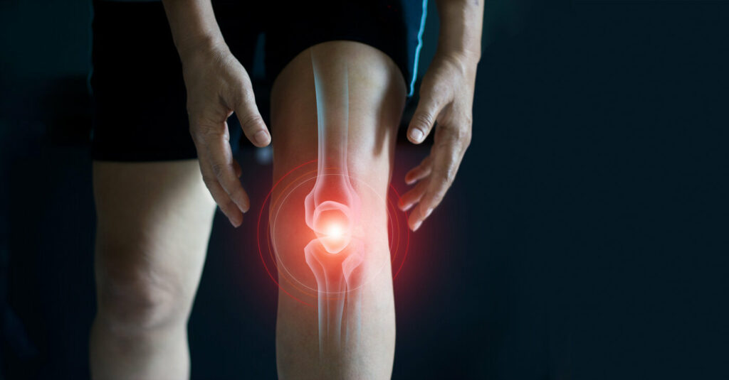
Knee
A recent 2020 study demonstrated that in 115 asymptomatic adults MRI scans showed abnormalities in the majority (97%) of knees.
> 30% of the knees had meniscus tears.
> 57% had cartilage abnormalities in the patellofemoral joint
> 48% had bone marrow abnormalities in the patellofemoral joint
> 19% had moderate cartilage lesions
> 31% had severe cartilage lesions
> 21% had moderate intensity tendon lesions
> 2% had partial ruptures of the ACL
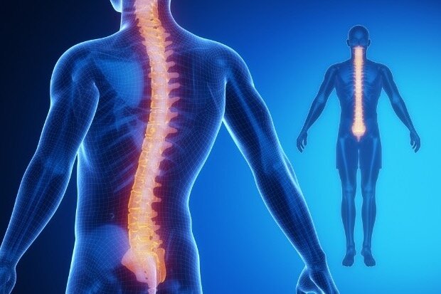
Spine
In the spine, the findings are similar, and normal aging features are found in people with no pain, no disability, and no symptoms (Wocial et.al 2021; Brinjikji et.al 2014) .
Imaging features Age groups (years)
20 30 40 50 60 70 80
Disc degeneration 37% 52% 68% 80% 88% 93% 96%
Disc signal loss 17% 33% 54% 73% 86% 94% 97%
Disc height loss 24% 34% 45% 56% 67% 76% 84%
Disc bulge 30% 40% 50% 60% 69% 77% 84%
Disc protrusion 29% 31% 33% 36% 38% 40% 43%
Annular fissure 19% 20% 22% 23% 25% 27% 29%
Facet degeneration 4% 9% 18% 32% 50% 69% 83%
Spondylolisthesis 3% 5% 8% 14%. 23% 35% 50%
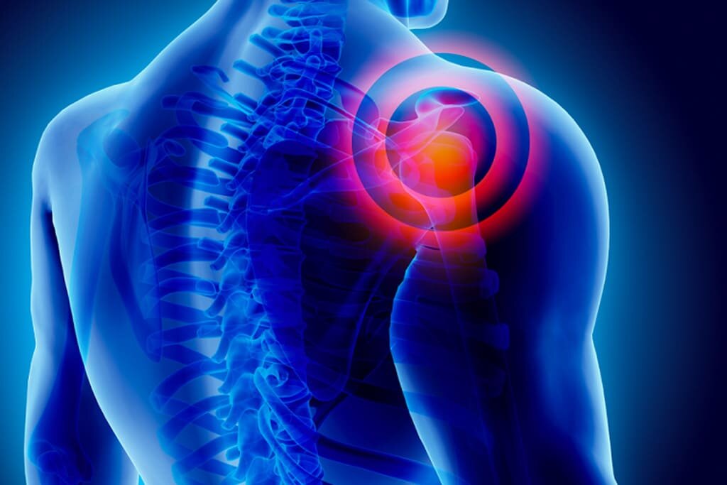
Shoulder
In the shoulder, the findings are similar, and normal aging features are found in people with no pain, no disability, and no symptoms.
One study from Grisih et.al 2011 demonstrated a staggering 96% of subjects with no pain or issues had at least one so-called shoulder pathology on their ultrasound scans.
Another study by Schwartzberg et.al 2016 demonstrated that 72% of middle-aged subjects had non-symptomatic superior labral lesions in their shoulder.
Liu et.al 2021 looked at the prevalence of shoulder pathology in young non-athletic asymptomatic subjects and demonstrated that 9% of 18-29-year-olds have labral abnormalities on MRI.
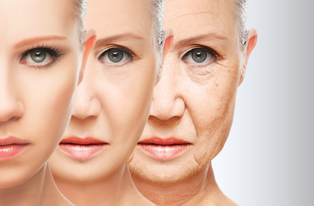
Aging On The Inside
We like to use this simple analogy of ageing on the inside. When our skin wrinkles, our hair greys, we can’t expect to look and feel the same inside. These processes of ageing occur every day that you are living and should be accepted as the norm. But what you don’t have to accept is a pain as you age. See our post on the ‘Ageing Athlete’ to understand more.
Conclusion:
Routine imaging is not necessary in most cases, but it can be extremely harmful. Imaging findings can in fact create unnecessary fear in patients, especially when clinicians don’t take the time to give proper context and explanations to the patient. Unfortunately we still often see clients whose radiological reports have been directly sent to them explaining their findings in medical jargon, with or without a dangerous form of language that is easily misinterpreted.
Always discuss your concerns and any questions you may have with your healthcare provider and remember a scan does not show pain it shows anatomy.
We are here for you
If you’re in pain and would like to talk to us about getting some help, some specialist advice, or if you are looking for a diagnosis, remember we are always here to help you.
Appointments remain limited and we are experiencing an exceptionally high demand for our physio services since UK restrictions were lifted, so please contact us immediately to avoid a long wait.
If you would like to get one of our limited slots, please click HERE to email or CALL us on 07900603617.
Horga, L.M., Hirschmann, A.C., Henckel, J., Fotiadou, A., Di Laura, A., Torlasco, C., D’Silva, A., Sharma, S., Moon, J.C. and Hart, A.J., 2020. Prevalence of abnormal findings in 230 knees of asymptomatic adults using 3.0 T MRI. Skeletal radiology, 49(7), pp.1099-1107
Brinjikji, W., Luetmer, P.H., Comstock, B., Bresnahan, B.W., Chen, L.E., Deyo, R.A., Halabi, S., Turner, J.A., Avins, A.L., James, K. and Wald, J.T., 2015. Systematic literature review of imaging features of spinal degeneration in asymptomatic populations. American Journal of Neuroradiology, 36(4), pp.811-816.
Wocial, K., Feldman, B.A., Mruk, B., Sklinda, K., Walecki, J. and Waśko, M., 2021. Imaging features of the aging spine. Polish Journal of Radiology, 86, p.e380.
Schwartzberg, R., Reuss, B.L., Burkhart, B.G., Butterfield, M., Wu, J.Y. and McLean, K.W., 2016. High prevalence of superior labral tears diagnosed by MRI in middle-aged patients with asymptomatic shoulders. Orthopaedic journal of sports medicine, 4(1), p.2325967115623212.


